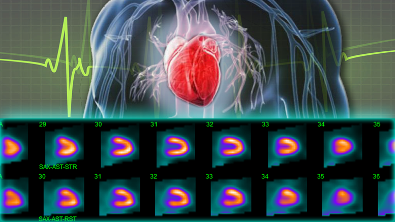
The heart supplies blood and oxygen to keep all the other systems functioning. When heart health is in question, specialized tests can help assess its performance under different conditions. One diagnostic tool is the nuclear stress test, a medical imaging procedure designed to test heart functions during physical activity and at rest. Explore the purpose of this procedure and who might need it:
Nuclear Stress Test Basics
This test is a non-invasive diagnostic procedure that uses a small amount of radioactive material to create images of the heart. These images allow healthcare providers to evaluate blood flow to the heart muscle. It helps them identify areas with reduced blood supply, and determine how well the heart pumps. The test is commonly performed in two phases.
The tests take place while the patient is at rest and another while the heart is undergoing physical exercise or medication that mimics the effects of exercise. Traditional stress tests only uses an electrocardiogram (ECG) to monitor heart activity. Nuclear stress tests provide more detailed visual information.
Test Candidates
A nuclear stress test is typically recommended for individuals showing symptoms that may indicate coronary artery disease or other heart-related conditions. Symptoms such as chest pain, shortness of breath, fatigue during physical activity, or irregular heartbeats prompt a healthcare provider to suggest this test as part of a broader diagnostic evaluation. It is frequently used for patients undergoing risk assessment before surgery, especially if they have risk factors for cardiovascular issues.
What Testing Detects
The main goal of this test is to detect issues related to blood flow to the heart. It can reveal whether parts of the heart are not receiving enough blood or if there are blockages in the coronary arteries. Additionally, it can identify areas of damage from a previous heart attack or assess conditions such as heart failure. By analyzing the images obtained during the test, cardiologists can determine if there are abnormalities in heart function.
Testing Process
The procedure typically begins with the placement of an intravenous (IV) line, through which a small amount of radioactive material is injected. This material, a radiotracer, travels to the heart and emits signals captured by a special camera. The test has two main phases:
- Resting Phase: Images are taken while the patient is at rest to establish a baseline for comparison.
- Stress Phase: During this phase, the patient either engages in physical exercise (like walking on a treadmill) or receives medication to simulate exercise. Additional images are taken afterward.
Test Results Explained
After the test, the images are analyzed to assess blood flow, identify blockages, and evaluate overall heart function. Areas that receive diminished blood flow during exercise but look normal at rest may indicate partial blockages. A cardiologist interprets these results and discusses what they mean for the patient’s health. Depending on the findings, further tests or treatments may be recommended to address any potential concerns.
Seek Care from a Cardiologist
The nuclear stress test is a helpful diagnostic tool that can provide valuable insights into heart health. Suppose you have symptoms or risk factors associated with heart disease. In that case, discussing options with a cardiologist may be an important step. Your cardiologist can provide tailored advice and explore treatment options to support your overall cardiovascular well-being.
